
Introduction
For all equine sports and disciplines, motion is a crucial element. We make big demands on how horses move, where they move, and how fast they move. Understanding the ‘equine motion muscles’ is valuable information for any horse person.
It is also valuable to see how equine motion muscles correlate to the muscles and structures of the human body. With the acknowledgement that horses mirror us on various levels, making sure your ‘motion muscles’ are conveying exactly what you’d like them to, can profoundly impact your performance.
Psychological, emotional, spiritual and physical components are indelibly intertwined. Studying equine anatomy and its relationship to our own can yield extraordinary insights, and can help us understand ourselves and our horses better.
Movement of a structured, almost immovable torso
Shown below, is a diagram of the horse. Included are muscles – mostly represented by lines, some with a transparent extension for simplicity – that are primarily involved with ‘initial’ motion.
Even though the flexor and extensor muscles of the forearm and gaskin areas contribute, they would not find their purpose in locomotion without the ability of the leg to swing forward and backward. It would be like standing still and lifting and lowering your foot without the notion of rotating your leg forward from the hip.
Once you’ve moved your leg forward, you’d have the opportunity to straighten your leg and reach forward with your foot. That’s why the swingers are illustrated here. Keep in mind; there are a lot more muscles in the horse that are designed to keep things ‘still’.
Many muscles around the torso for example, are designed for support and protection, not propulsion. Muscles of the spine are designed to prevent mobility for obvious reasons.
Note: There is no specificity in the horses anatomy for carrying weight. There aren’t an extra set of legs under the burden of a rider, nor are there slings carrying the torso from an imaginary point in the sky.
Over-extension and/or fatigue of these muscles increases the risk of serious injury, and under normal circumstances, all these muscles work in excellent harmony. Nature has provided the horse with a magnificent package…
Equine Movement Muscles
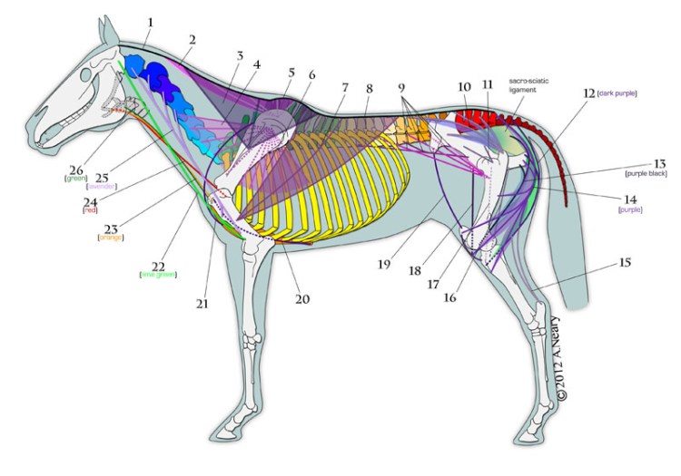
Here’s the basic rundown of the ‘equine motion muscles’ (and a couple of key ligaments), along with their names and basic functions:
1. Nuchal ligament: continuation of supraspinatus ligament. Attaches to rear of cranium or skull.
2. Cervical portion of Rhomboid muscle: scapular (scapula = shoulder blade) rotation, pulls head up when leg is fixed – pulls neck to one side
3. Trapezius: thoracic and cervical portions involved in scapular rotation
Ventral Serrate (#4 and #6): forms a sling, hanging the trunk from the scapula – raises thorax – unilaterally shifts weight to one side
4. Cervical ventral serrate: scapular rotation – pulls upper end of scapula cranially and ventrally, raises neck when leg is fixed, or pulls neck to one side
5. Thoracic portion of Rhomboid muscle: scapular rotation
6. Thoracic ventral serrate: scapular rotation, pulls upper end of scapula caudally and ventrally -possibility assist in breathing ‘in’
Note: The horse does not have collar bones, and therefore, the trunk is suspended in-between the shoulder blades by muscles. Any and all training and exercise methods that ignore the proper development of these muscles – especially under the weight of the rider, the equipment, and the restrictions caused by the handling of the reins – cause the entire spine and its neighboring apparatus to sink and become compressed. This limits the true, natural, innate, inborn motility and freedom of the horse. The horse’s body (and mind) is forced to compensate, which over time will reveal itself in mobility issues, lameness issues and other ‘idiopathic’ problems.
7. Latissimus dorsi: pulls leg back, moves trunk forward over advanced/fixed leg
8. Supraspinatus ligament: connected to all spinous processes of thoracic and lumbar vertebrae
9. PSOAS group
a. Psoas major: flexes hip, rotates thigh laterally – swinging stifle outward during motion
b. Psoas minor: flexes or fixes lumbar and lumbosacral joints; inclines pelvis laterally on loins
c. Ilio-psoas (pulls leg forward)
d. Iliacus component
10. Middle Gluteal: pulls leg back as in kicking – moves trunk forward over fixed leg in conjunction with latissimus dorsi; contributes to rearing and jumping
11. Deep Gluteal: pulls leg back and rotates stifle inward (rotate thigh medially)
All parts of the Biceps Femoris, Semitendinosus, and Semimenbranosis (#12, #13, #14) are collectively called the hamstring muscles. They extend the hip, stifles and hock in bringing the leg back during forward movement. They are also responsible for the action of rearing, kicking and jumping.
12. Semitendinosus: extend hip and hock, flex stifle and rotate thigh medially during backwards motion of the leg
13. Semimenbranosus: extends hip, adducts leg
14. Biceps Femoris: extends hip, stifle and hock in leg retraction and locomotion
a. Cranial (toward head) part of Biceps Femoris: exends hip and stifle
b. Middle part of Biceps Femoris: extends hip
c. Caudal (toward tail) part of Biceps Femoris: flexes stifle, extends hock
15. Accessary tarsal tendons: extend hock
16. Gracilis: adducts leg and extends stifle and hock
17. Adductor: adducts and retracts protracted limb
18. Sartorius: flexes hip, adducts leg
19. Tensor muscle of lateral femoral fascia: flexes hip, extends stifle, tightens femoral fascia
20. Ascending Pectoral: retracts leg from forward position, draws trunk forward over advanced leg
21. Subclavius (pre-scapular part of deep pectoral): similar to ascending pectoral, assists ventral serrate in suspending the trunk
22. Brachiocephalic: pulls leg forward, lowers head and extends neck when leg in fixed position. Can move neck to one side.
23. Sternohyoid: pulls tongue and larynx back and down in swallowing; fix hyoid when tongue muscles act
24. Omohyoid: pulls hyoid and root of tongue toward tail
25. Omotransverse: similar to Brachiocephalic
26. Sternomandibular: bilaterally pulls head and neck down; unilaterally flexes head and neck to the side
Here’s another example for the application of this information. During my time as a coach and instructor, this was invaluable…
Many riders, especially dressage riders, encounter the infamous ‘bulging shoulder’ at one time or another. You now have the knowledge and power to think about how you might use your own brachiocephalicus and thorax muscles to help make corrections. This includes your breath and breathing habits, which greatly affect your fluidity and performance, and therefore, affect your horse’s fluidity and performance…their capacity for mirroring every aspect of you, is awake, alive, humbling and astonishing…

One thought on “Equine Motion Muscles”